Arteries In Neck Labeled / Teach Besides Me: Pictures Of Veins And Arteries - The oa then turns medially, giving off 1 to 5 posterior ciliary arteries (pca) that subsequently branch into the long and short posterior ciliary arteries (lpca and spca respectively) which perforate the sclera posteriorly in the vicinity of the optic nerve and macula to supply the posterior uveal tract.
Arteries In Neck Labeled / Teach Besides Me: Pictures Of Veins And Arteries - The oa then turns medially, giving off 1 to 5 posterior ciliary arteries (pca) that subsequently branch into the long and short posterior ciliary arteries (lpca and spca respectively) which perforate the sclera posteriorly in the vicinity of the optic nerve and macula to supply the posterior uveal tract.. Learn these parts of body names to increase your vocabulary words in This anatomy section promotes the use of the terminologia anatomica, the i. Ct of the head and neck Superficial dissection of the right side of the neck, showing the carotid and subclavian arteries. May 06, 2021 · this head and neck anatomy atlas is an educational tool for studying the normal anatomy of the face based on a contrast enhanced multidetector computed tomography imaging (axial and coronal planes).
May 06, 2021 · this head and neck anatomy atlas is an educational tool for studying the normal anatomy of the face based on a contrast enhanced multidetector computed tomography imaging (axial and coronal planes). May 06, 2021 · the arteries and veins of the face and neck were labeled focusing on large trunks emerging from the external carotid artery or draining into the jugular vein system. Superficial dissection of the right side of the neck, showing the carotid and subclavian arteries. Cartilaginous joints are a type of joint where the bones are entirely joined by cartilage, either hyaline cartilage or fibrocartilage. Ct of the head and neck
Vascular System Models - Arteries, Veins, Blood Cells ... from i.pinimg.com Superficial dissection of the right side of the neck, showing the carotid and subclavian arteries. Branch of vertebral artery and thyrocervical trunk is labeled. May 06, 2021 · the arteries and veins of the face and neck were labeled focusing on large trunks emerging from the external carotid artery or draining into the jugular vein system. The oa then turns medially, giving off 1 to 5 posterior ciliary arteries (pca) that subsequently branch into the long and short posterior ciliary arteries (lpca and spca respectively) which perforate the sclera posteriorly in the vicinity of the optic nerve and macula to supply the posterior uveal tract. May 06, 2021 · this head and neck anatomy atlas is an educational tool for studying the normal anatomy of the face based on a contrast enhanced multidetector computed tomography imaging (axial and coronal planes). These joints generally allow more movement than fibrous joints but less movement than synovial joints. The cervical spine has 7 stacked bones called vertebrae, labeled c1 through c7. Different parts of the body in english with body parts pictures and examples.
These joints generally allow more movement than fibrous joints but less movement than synovial joints.
Ct of the head and neck Branch of vertebral artery and thyrocervical trunk is labeled. This anatomy section promotes the use of the terminologia anatomica, the i. The neck is connected to the upper back through a series of seven vertebral segments. The common carotid arteries (c) are normal but the right internal jugular vein (j) is enlarged with a dense or enhancing wall surrounding the more lucent intraluminal clot (arrow). The top of the cervical spine connects to the skull, and the bottom connects to the upper back at about shoulder level. Superficial dissection of the right side of the neck, showing the carotid and subclavian arteries. Neuroanatomy encompasses the anatomy of all structures of the central nervous system (cns), which includes the brain and the spinal cord, and their supporting structures. Internal thoracic artery branches from same segment, but inferiorily, and is therefore not visible. The cervical spine has 7 stacked bones called vertebrae, labeled c1 through c7. May 06, 2021 · this head and neck anatomy atlas is an educational tool for studying the normal anatomy of the face based on a contrast enhanced multidetector computed tomography imaging (axial and coronal planes). May 06, 2021 · the arteries and veins of the face and neck were labeled focusing on large trunks emerging from the external carotid artery or draining into the jugular vein system. Second year medical clerkship in anatomy.
Cartilaginous joints are a type of joint where the bones are entirely joined by cartilage, either hyaline cartilage or fibrocartilage. The neck is connected to the upper back through a series of seven vertebral segments. The cervical spine has 7 stacked bones called vertebrae, labeled c1 through c7. Branch of vertebral artery and thyrocervical trunk is labeled. Internal thoracic artery branches from same segment, but inferiorily, and is therefore not visible.
Head and neck anatomy - Wikipedia from upload.wikimedia.org The oa then turns medially, giving off 1 to 5 posterior ciliary arteries (pca) that subsequently branch into the long and short posterior ciliary arteries (lpca and spca respectively) which perforate the sclera posteriorly in the vicinity of the optic nerve and macula to supply the posterior uveal tract. Internal thoracic artery branches from same segment, but inferiorily, and is therefore not visible. The top of the cervical spine connects to the skull, and the bottom connects to the upper back at about shoulder level. Neuroanatomy encompasses the anatomy of all structures of the central nervous system (cns), which includes the brain and the spinal cord, and their supporting structures. Learn these parts of body names to increase your vocabulary words in This anatomy section promotes the use of the terminologia anatomica, the i. The neck is connected to the upper back through a series of seven vertebral segments. The cervical spine has 7 stacked bones called vertebrae, labeled c1 through c7.
The common carotid arteries (c) are normal but the right internal jugular vein (j) is enlarged with a dense or enhancing wall surrounding the more lucent intraluminal clot (arrow).
The oa then turns medially, giving off 1 to 5 posterior ciliary arteries (pca) that subsequently branch into the long and short posterior ciliary arteries (lpca and spca respectively) which perforate the sclera posteriorly in the vicinity of the optic nerve and macula to supply the posterior uveal tract. Learn these parts of body names to increase your vocabulary words in May 06, 2021 · the arteries and veins of the face and neck were labeled focusing on large trunks emerging from the external carotid artery or draining into the jugular vein system. Branch of vertebral artery and thyrocervical trunk is labeled. The cervical spine has 7 stacked bones called vertebrae, labeled c1 through c7. These joints generally allow more movement than fibrous joints but less movement than synovial joints. Ct of the head and neck Branch of vertebral artery and thyrocervical trunk is labeled. Superficial dissection of the right side of the neck, showing the carotid and subclavian arteries. Internal thoracic artery branches from same segment, but inferiorily, and is therefore not visible. Second year medical clerkship in anatomy. May 06, 2021 · this head and neck anatomy atlas is an educational tool for studying the normal anatomy of the face based on a contrast enhanced multidetector computed tomography imaging (axial and coronal planes). The common carotid arteries (c) are normal but the right internal jugular vein (j) is enlarged with a dense or enhancing wall surrounding the more lucent intraluminal clot (arrow).
Cartilaginous joints are a type of joint where the bones are entirely joined by cartilage, either hyaline cartilage or fibrocartilage. Second year medical clerkship in anatomy. Most cases resolve without sequelae with early and appropriate treatment. Internal thoracic artery branches from same segment, but inferiorily, and is therefore not visible. Superficial dissection of the right side of the neck, showing the carotid and subclavian arteries.
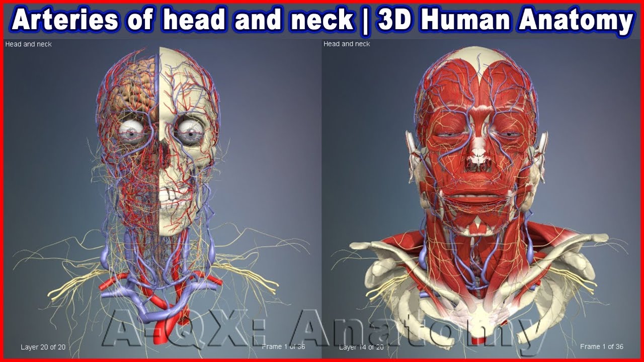
Arteries of head and neck | 3D Human Anatomy | Organs ... from i.ytimg.com Most cases resolve without sequelae with early and appropriate treatment. Superficial dissection of the right side of the neck, showing the carotid and subclavian arteries. Branch of vertebral artery and thyrocervical trunk is labeled. Learn these parts of body names to increase your vocabulary words in Different parts of the body in english with body parts pictures and examples. Interactive labeled images allow a comprehensive study of the anatomical structures. The common carotid arteries (c) are normal but the right internal jugular vein (j) is enlarged with a dense or enhancing wall surrounding the more lucent intraluminal clot (arrow). Second year medical clerkship in anatomy.
The cervical spine has 7 stacked bones called vertebrae, labeled c1 through c7.
May 06, 2021 · this head and neck anatomy atlas is an educational tool for studying the normal anatomy of the face based on a contrast enhanced multidetector computed tomography imaging (axial and coronal planes). Second year medical clerkship in anatomy. Superficial dissection of the right side of the neck, showing the carotid and subclavian arteries. Branch of vertebral artery and thyrocervical trunk is labeled. Cartilaginous joints are a type of joint where the bones are entirely joined by cartilage, either hyaline cartilage or fibrocartilage. Different parts of the body in english with body parts pictures and examples. Ct of the head and neck The oa then turns medially, giving off 1 to 5 posterior ciliary arteries (pca) that subsequently branch into the long and short posterior ciliary arteries (lpca and spca respectively) which perforate the sclera posteriorly in the vicinity of the optic nerve and macula to supply the posterior uveal tract. Superficial dissection of the right side of the neck, showing the carotid and subclavian arteries. Interactive labeled images allow a comprehensive study of the anatomical structures. These joints generally allow more movement than fibrous joints but less movement than synovial joints. Most cases resolve without sequelae with early and appropriate treatment. Learn these parts of body names to increase your vocabulary words in
Superficial dissection of the right side of the neck, showing the carotid and subclavian arteries arteries in neck. Different parts of the body in english with body parts pictures and examples.


0 Komentar